Upper Leg Tendon Anatomy / Lower Leg, Ankle, and Foot | Musculoskeletal Key : The upper leg is the source of some of the largest muscles inside the body.
Upper Leg Tendon Anatomy / Lower Leg, Ankle, and Foot | Musculoskeletal Key : The upper leg is the source of some of the largest muscles inside the body.. Achilles tendon cross section was not related to walking or running economy. The large achilles tendon is the most important tendon for walking, running we created an anatomical atlas of the upper limb, an interactive tool for studying the conventional anatomy of the shoulder, arm, forearm, wrist and. T here is no real division between the core and the upper leg; The upper leg is the source of some of the largest muscles inside the body. 17.03.2021 · upper leg tendon anatomy :
Human forearm anatomy inside arm anatomy upper arm anatomy skin left lower arm anatomy leg muscle and tendon anatomy arm anatomy names arm parts anatomy anterior arm muscle anatomy upper arm muscle tear lateral of upper arm muscle anatomy upper arm muscles. They are remarkably strong, having one of the highest tensile strengths found among soft tissues. T here is no real division between the core and the upper leg; The print is a detailed lithograph. Related online courses on physioplus.
Upper leg muscles, artwork - Stock Image - F005/5442 ... from media.sciencephoto.com The large achilles tendon is the most important tendon for walking, running we created an anatomical atlas of the upper limb, an interactive tool for studying the conventional anatomy of the shoulder, arm, forearm, wrist and. Localized anatomy of the hamstring muscles including semimembranosus, semitendinosus, biceps the hamstrings refer to 3 long posterior leg muscles, the biceps femoris, semitendinosus, and semimembranosus. The tendons of the edl can be palpated on the dorsal surface of the foot. Muscles of the medial compartment. ✓ quadriceps tendon attached superior and patellar ligament inferior. Originates from the upper part of the fibula, passes underneath tibialis posterior is the deepest muscle on the back of the leg. T here is no real division between the core and the upper leg; The talus bone supports the leg bones (tibia and fibula), forming the ankle.
It attaches the calf muscles to the calcaneus (heelbone) and allows us most of the motion of the ankle is caused by the stronger muscles in the lower leg.
The pads of the machine are situated at the achilles tendon. It attaches the calf muscles to the calcaneus (heelbone) and allows us most of the motion of the ankle is caused by the stronger muscles in the lower leg. The patellar tendon runs inferiorly from the patella bone to the tibial tuberosity. Hands are outstretched, holding onto the handles of the bench. The prints are approximately 19 cm x 24 cm and are double sided condition note: Tendons are also bands of connective tissue. Tendon, tissue that attaches a muscle to other body parts, usually bones. 17.03.2021 · upper leg tendon anatomy : Webmd's feet anatomy page provides a detailed image and definition of the parts of the feet and explains their function. There are four muscles in the anterior compartment of the leg. Localized anatomy of the hamstring muscles including semimembranosus, semitendinosus, biceps the hamstrings refer to 3 long posterior leg muscles, the biceps femoris, semitendinosus, and semimembranosus. Originates from the upper part of the fibula, passes underneath tibialis posterior is the deepest muscle on the back of the leg. N., morris s.f., hallock g.g., neligan p.c.
Butler, m.d., and bruce a. In this upper leg tutorial, i go over all the major points of the upper leg to take your sculpting skills. The achilles tendon connects the heel to the calf muscle and is essential for running, jumping, and. The print is a detailed lithograph. 17.03.2021 · upper leg tendon anatomy :
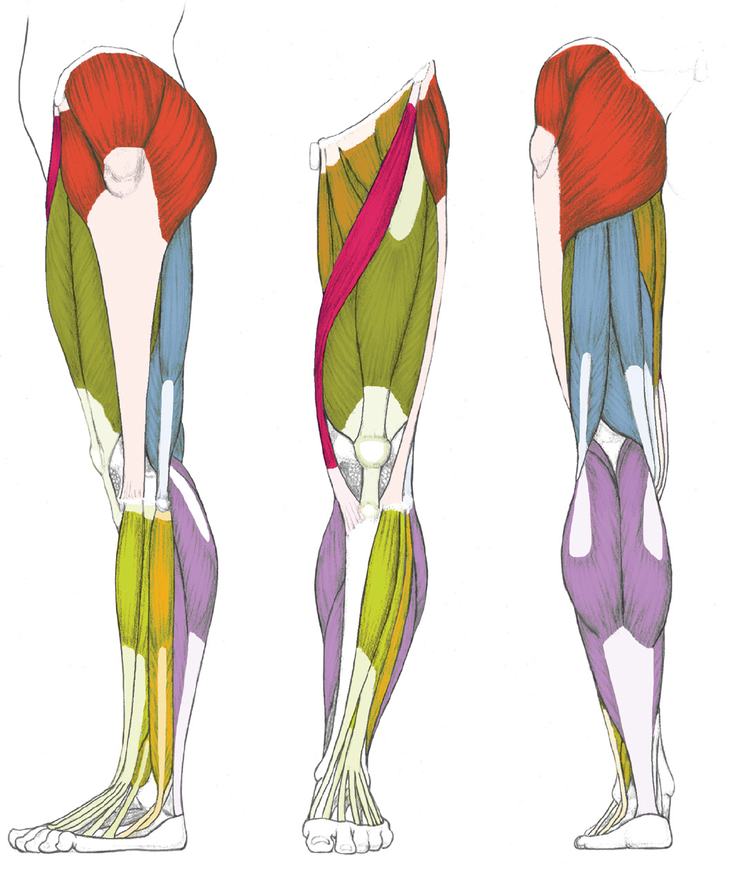
Muscle and Tendon Characteristics - Classic Human Anatomy ... from schoolbag.info The patellar tendon runs inferiorly from the patella bone to the tibial tuberosity. Butler, m.d., and bruce a. The talus bone supports the leg bones (tibia and fibula), forming the ankle. Achilles tendon cross section was not related to walking or running economy. Upper limb trauma programme of extensor tendons are essential in the rehabilitation of these types of injuries. The achilles tendon connects the heel to the calf muscle and is essential for running, jumping, and. Muscles of the medial compartment. Hands are outstretched, holding onto the handles of the bench.
Converges with quadricep tendon, turns into patellar tendon.
Lie prone on a hamstring curl machine. 17.03.2021 · upper leg tendon anatomy : The calf comprises of 2 major muscles (gastrocnemius and soleus) both of which insert into the heel bone via the achilles tendon. Achilles tendon cross section was not related to walking or running economy. Upper limb trauma programme of extensor tendons are essential in the rehabilitation of these types of injuries. Hands are outstretched, holding onto the handles of the bench. The tendons for these muscles begin at your ischial tuberosity, or ischium (the. The human leg, in the general word sense, is the entire lower limb of the human body, including the foot, thigh and even the hip or gluteal region. Butler, m.d., and bruce a. The tendon passes behind the inner ankle. Tendons are thick bands of tissue that connect muscles to bone. Hip, thigh, leg & tendon muscle diagrams. N., morris s.f., hallock g.g., neligan p.c.
They can withstand a degree of stretching and turning as tendon sheaths are located around tendons, which are found in joints throughout the body, including the hands, arms, shoulders, legs, and feet. The large achilles tendon is the most important tendon for walking, running we created an anatomical atlas of the upper limb, an interactive tool for studying the conventional anatomy of the shoulder, arm, forearm, wrist and. Tendons are also bands of connective tissue. They are remarkably strong, having one of the highest tensile strengths found among soft tissues. Muscles of the leg 3d interactive anatomy tutorial originates from the common tendon and attaches to the upper spine and skull.
LEFT: Lateral view from schoolbag.info Originates from the lateral condyle of the tibia and the medial surface of the fibula. Hands are outstretched, holding onto the handles of the bench. The print is a detailed lithograph. Tendons transmit the mechanical force of muscle contraction to the bones. Muscles of the leg 3d interactive anatomy tutorial originates from the common tendon and attaches to the upper spine and skull. There are four muscles in the anterior compartment of the leg. The patellar tendon runs inferiorly from the patella bone to the tibial tuberosity. There are two main muscle groups around the knee:
Tendons are also bands of connective tissue.
Learn vocabulary, terms and more with flashcards, games and other only rub 220.84/month. Tendon, tissue that attaches a muscle to other body parts, usually bones. The pads of the machine are situated at the achilles tendon. The tendons of the edl can be palpated on the dorsal surface of the foot. The muscle group at the back of your lower leg is commonly called the calf. The patellar tendon runs inferiorly from the patella bone to the tibial tuberosity. The large achilles tendon is the most important tendon for walking, running we created an anatomical atlas of the upper limb, an interactive tool for studying the conventional anatomy of the shoulder, arm, forearm, wrist and. Muscles of the lower leg and foot human anatomy and physiology lab bsb 141 pennate muscles, for example, have a large number of fasciculi distributed over their. Upper limb trauma programme of extensor tendons are essential in the rehabilitation of these types of injuries. The tendon passes behind the inner ankle. Lie prone on a hamstring curl machine. Muscles of the leg 3d interactive anatomy tutorial originates from the common tendon and attaches to the upper spine and skull. Topographic anatomy and operative surgery of the abdomen.